A research team at Wuyi University in China has developed a bionic finger that can recognize internal shapes and textures of complex layered objects. It scans objects by applying pressure to the surface, detecting both external and internal structures as it moves along. The finger measures the degree of compression of a surface, which provides information about the relative softness or stiffness of the object being touched. The device has been tested on complex objects such as a simulated human skeleton, and it can achieve a spatial resolution of at least 500 µm in the x and y planes and 200 µm in the z-axis direction. The researchers hope to incorporate the bionic finger into robots or prosthetics for use in biomedical and robotic engineering. Future clinical applications could include helping physicians diagnose lumps under the skin, such as those caused by breast cancer lesions, and non-invasive industrial or research testing.
A Revolutionary ‘Bionic Finger’ That Can Create 3D Maps of Human Tissues
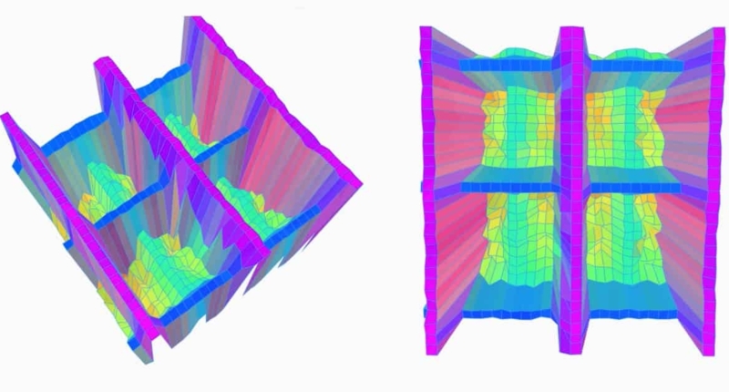
A research team at Wuyi University in China has developed a revolutionary ‘bionic finger’ that can create 3D maps of complex human tissues and flexible electronics. By using tactile tomography, the finger can recognize the internal shapes and textures of complex layered objects by simply touching their exterior surfaces. The data is then transmitted to a computer to generate 3D maps.
This innovative development could pave the way for the use of smart bionic fingers in diagnostic imaging, as an alternative or complementary method to ultrasound or X-ray exams. The technology’s inspiration comes from the human finger, which has the most sensitive tactile perception known to man.
The team explains that when the skin of a human finger touches an object, it undergoes mechanical deformation such as compression, stretching, or drag. These deformations stimulate mechanoreceptors to emit electrical impulses. The electrical impulses travel through the central nervous system to the somatosensory cortex of the brain and are finally integrated by the brain to recognize the characteristics of the material.
To replicate this process, the team designed the bionic finger using carbon fiber beams as mechanoreceptors. The finger consists of a bundle of carbon fibers with a 0.5 mm-diameter metal cylinder mounted on top as the contact tip. The fibers connect to a signal processing module, which contains signal acquisition and controller modules that combine with the sensor to establish a tactile feedback system.
The finger scans an object by applying pressure to the surface, detecting both external and internal structures as it moves along. It measures the degree of compression of a surface, which provides information about the relative softness or stiffness of the object being touched. The bionic finger can achieve a spatial resolution of at least 500 µm in the x and y planes and 200 µm in the z-axis direction.
To test the finger’s capabilities, the research team performed a series of investigations on complex objects. One of the tests involved recognizing a rigid letter “A” buried beneath a soft silicon outer layer. They also tested the finger with a simulated human skeleton comprising a soft silicon “skin” layer, a “muscle” layer, a layer containing simulated blood vessels, and hard polymer skeletal “bones.”
In conclusion, the smart bionic finger’s ability to create 3D maps of complex human tissues and flexible electronics is groundbreaking. Its future use in diagnostic imaging could be a game-changer for the medical industry, especially as it provides a non-invasive alternative or supplement to traditional methods such as ultrasound or X-ray exams. The possibilities of this technology are limitless and could lead to significant advancements in various fields, including medicine, robotics, and engineering.
A Revolutionary Bionic Finger that can Diagnose Tissue and Electronic Problems
The bionic finger, developed by researchers at Wuyi University, accurately reproduces the tissue structure and can locate simulated blood vessels beneath the muscle layer. However, the researchers advise that improvements are needed to reconstruct blood vessels with greater precision and to enable the finger to recognize more complex 3D structures.
In addition to tissue diagnosis, the researchers also investigated the bionic finger’s ability to diagnose problems in electronic devices. After scanning the surface of an encapsulated flexible circuit system, the finger created a 3D map of its internal electrical components. It accurately located where the circuit was disconnected and identified a mis-drilled hole without breaking through the encapsulating layer.
The researchers aim to incorporate the bionic finger into robots or prosthetics for use in biomedical and robotic engineering. Future clinical applications could include using the finger to help physicians diagnose lumps under the skin, such as those caused by breast cancer lesions. They anticipate that it will also be an excellent tool for non-invasive industrial or research testing.
Don’t miss interesting posts on Famousbio

