


A recent study recommends redefining prostate bed contouring guidelines for salvage radiation therapy to improve outcomes for prostate cancer patients. The study, which used PSMA PET to image patterns of prostate bed recurrence, found that current contouring guidelines based on expert consensus miss a significant number of cancer lesions and may irradiate healthy tissues unnecessarily. Prostate cancer is one of the most common types of cancer among men, and approximately one-third of patients who undergo radical prostatectomy experience disease progression within ten years. Salvage radiation therapy is a potentially curative treatment option for these patients, but the study found that current guidelines do not take advantage of the information available from novel imaging techniques such as PSMA PET. The authors recommend redefining prostate bed contouring guidelines for SRT to improve outcomes for patients receiving radiotherapy post-radical surgery. Nuclear medicine and molecular imaging play a crucial role in guiding decision-making for cancer treatment, and the study highlights their growing importance in individualized, tailored treatments for patients with prostate cancer.
PSMA PET Mapping Reveals Inadequacy of Current Radiotherapy Contouring Guidelines for Prostate Cancer Treatment

A new study published in The Journal of Nuclear Medicine reveals that current radiotherapy contouring guidelines used to determine target areas for prostate cancer treatment are inadequate, as they miss a significant number of cancer lesions and may cause unnecessary irradiation of healthy tissues. The study calls for the redefinition of prostate bed contouring guidelines to improve outcomes for patients.
One-third of prostate cancer patients who undergo radical prostatectomy experience disease progression within 10 years, making salvage radiation therapy (SRT) a potentially curative treatment option. However, SRT currently follows contouring guidelines based on expert consensus, which do not incorporate the information available from novel imaging techniques like PSMA PET.
PSMA PET is one of the most accurate methods for detecting tumor recurrence after prostatectomy and has changed patterns of care for prostate cancer patients since its FDA approval in December 2020. The researchers sought to analyze the patterns of prostate bed recurrence with PSMA PET and see how they compared to current expert consensus contouring guidelines from the Radiation Therapy Oncology Group (RTOG).
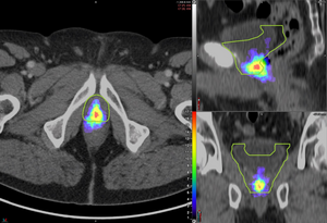
The study involved 127 prostate cancer patients experiencing PSA persistence or biochemical recurrence after radical prostatectomy. The researchers used PSMA PET to image the patterns of prostate bed recurrence and assessed the location of recurrent lesions for each scan. They then determined the clinical target volume (CTV) based on the RTOG contouring guidelines and categorized the lesions as completely covered, partially covered, or not covered by the guidelines.
The researchers found that the recurrent lesions were completely covered in the CTV in 53 percent of patients, partially covered in 34 percent of patients, and not covered in 13 percent of patients. Moreover, some areas in the CTV had no evidence of lesions at all. Therefore, the study suggests that the current radiotherapy contouring guidelines miss a significant number of lesions, and as a result, there is a need to redefine prostate bed contouring guidelines to improve outcomes for patients.
In conclusion, the study highlights the inadequacy of current radiotherapy contouring guidelines for prostate cancer treatment and emphasizes the importance of incorporating the information available from novel imaging techniques like PSMA PET in the treatment plan to improve patient outcomes.
PSMA PET/CT Atlas Can Help Redefine Prostate Bed Contouring Guidelines for Salvage Radiation Therapy
A new study titled “PSMA PET/CT–based Atlas for Prostatic Bed Recurrence after Radical Prostatectomy: Clinical Implications for Salvage Radiation Therapy Contouring Guidelines” highlights the crucial role that nuclear medicine and molecular imaging play in guiding decision-making for cancer treatment, particularly in patients with prostate cancer.
The study, which was made available online in February 2023, recommends that current radiotherapy contouring guidelines used to determine target areas for prostate cancer treatment be redefined to improve outcomes for patients. According to the study, current contouring guidelines miss a significant number of cancer lesions and may cause unnecessary irradiation of healthy tissues.
The researchers used PSMA PET to image the patterns of prostate bed recurrence in 127 prostate cancer patients experiencing PSA persistence or biochemical recurrence after radical prostatectomy. They assessed the location of recurrent lesions for each PSMA PET scan and determined the clinical target volume (CTV) based on the Radiation Therapy Oncology Group contouring guidelines.
The study found that the recurrent lesions were completely covered in the CTV in 53 percent of patients, partially covered in 34 percent of patients, and not covered in 13 percent of patients. Therefore, the authors of the study recommend redefining prostate bed contouring guidelines for salvage radiation therapy (SRT) to improve the outcomes of patients receiving radiotherapy post-radical surgery.
According to Ida Sonni, MD, nuclear medicine physician and academic researcher in the Department of Radiological Sciences at the University of California, Los Angeles, “Our work confirms the crucial and growing role that nuclear medicine and molecular imaging have in guiding decision-making for cancer treatment. Nuclear medicine plays an essential part in the multidisciplinary management of patients with prostate cancer and facilitates the use of individualized, tailored treatments, which ultimately benefit all our patients.”
The authors of the study include researchers from the Ahmanson Translational Theranostics Division, Department of Molecular and Medical Pharmacology, David Geffen School of Medicine, University of California Los Angeles, Los Angeles, California, Department of Radiology, David Geffen School of Medicine, University of California, Los Angeles, California, and Department of Experimental and Clinical Medicine, Nuclear Medicine Unit, Magna Graecia University, Catanzaro, Italy.
About JNM and SNMMI
The Journal of Nuclear Medicine (JNM) is the world’s leading nuclear medicine, molecular imaging, and theranostics journal. JNM is published by the Society of Nuclear Medicine and Molecular Imaging (SNMMI), an international scientific and medical organization dedicated to advancing nuclear medicine and molecular imaging. The SNMMI is committed to precision medicine, which allows diagnosis and treatment to be tailored to individual patients, resulting in the best possible outcomes. Practitioners around the world access the journal 15 million times each year, providing them with the information they need to advance this rapidly expanding field.
Readers can access current and past issues of JNM online at http://jnm.snmjournals.org. For more information about SNMMI, visit www.snmmi.org.
Please note that AAAS and EurekAlert! are not responsible for the accuracy of news releases posted to EurekAlert! by contributing institutions or for the use of any information through the EurekAlert system.
Don’t miss interesting posts on Famousbio


