


Researchers have discovered that exosomes – small extracellular vesicles – vary by age, sex, and the extent of amyloid deposition in the brain. Scientists used a method called microdialysis to sample extracellular vesicles from the interstitial fluid of the hippocampus in living mice. The researchers found that as male wild-type or APP/PS1 mice aged, the concentration of EVs within their hippocampal interstitial fluid increased, whereas the concentration of EVs in female mice held steady, regardless of age or genotype. Microglia-derived EVs were found to be more responsive biomarkers to AD pathogenesis than neuron-derived EVs. The study showed that immune proteins were elevated in exosomes from 3-month-old APP/PS1 mice compared to young wild-type mice, but then did not increase later on. The findings could provide distinct signatures of extracellular vesicles at the site of AD pathology to look for in cerebrospinal fluid or blood of people with Alzheimer’s.
Exosomes: Potential Biomarkers for Brain Conditions
Extracellular vesicles (EVs) carrying a mix of RNAs, proteins, and metabolites reflect the type and state of their parent cell, offering a potential trove of biomarkers for brain conditions. Exosomes are a tiny type of EV that originate from endosomes that fuse with the plasma membrane. While brain-derived exosomes have been found in the cerebrospinal fluid and plasma, it has been challenging to determine how faithfully they reflect ongoing events within different parts of the brain.
Scientists have recently developed a method to intercept these vesicular packages within a living mouse, sampling EVs from the interstitial fluid of the hippocampus using microdialysis. Researchers from Wake Forest School of Medicine led by Gagan Deep and Shannon Macauley have used this method to examine the proteomes of microglial exosomes, a type of EV, in male and female amyloid models. They discovered that the protein contents of the vesicular packets varied markedly by age, sex, and the extent of amyloid deposition, and that exosomal contents are tied to microglial function, suggesting that they might be useful as disease biomarkers.


The findings reveal that hippocampal EVs increased dramatically with age in male mice but not in females. Moreover, female mice accumulated twice as much amyloid as their male counterparts, yet their microglia appeared to release fewer EVs containing fewer proteins. These differences suggest that exosomes from male and female mice respond differently to amyloid and that the proteomes of microglial exosomes differ between sexes in amyloid models.

Overall, this research demonstrates the potential for using exosomes as biomarkers for brain conditions and the importance of considering sex differences in disease studies.

A New Method for Studying Brain Communication
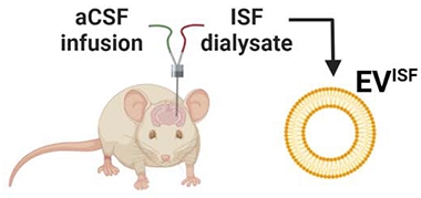
Scientists are using a new method called the vivo microdialysis to study how exosomes in the hippocampus relate to age, sex, and amyloidosis. The technique has previously been used to study the ebb and flow of proteins within brain fluid. Researchers inserted a guide cannula directly into the left hippocampus of living mice, through which they inserted a probe equipped with a 1,000kDa cut-off filter. Pumping in artificial cerebrospinal fluid, interstitial fluid (ISF) was slowly collected every 90 minutes over a period of 72 hours as the mice roamed freely. Eventually, enough fluid was collected to pellet small extracellular vesicles (EVs), including exosomes, via ultracentrifugation. The researchers then used flow cytometry and mass spectrometry to measure the number, size, and proteomes of the EVs.
Sex-Specific Differences in EV Concentration
As male wild-type or APP/PS1 mice aged, the concentration of EVs within their hippocampal ISF increased by about two orders of magnitude. For females, the concentration of EVs in the ISF held steady, regardless of age or genotype. Interestingly, by 9 months, female APP/PS1 mice had around double the load of amyloid plaques as males. “We were very surprised to see such a strong sex-specific difference in EV concentration with age,” said Shannon Macauley of Wake Forest School of Medicine.
Proteins within EVs Change with Age
For mice of both sexes and genotypes, the concentration of proteins within their EVs increased with age. Proteomic analysis of ISF EVs identified 436 proteins involved in numerous biological processes. Of these, 168 were shared across mice of both genotypes and both ages, pointing to a core group of hippocampal EV proteins. Smaller numbers of proteins were unique to each genotype and age. However, the concentration of total proteins increased with age regardless of genotype, but the diversity of proteins dropped with older age and APP/PS1 genotype. The number of biological processes in which the EV proteins were involved was also lower in APP/PS1 mice relative to wild-type animals.
This research sheds light on the potential of exosomes as a biomarker for brain conditions and the importance of considering sex differences in disease studies. It also highlights the potential of the vivo microdialysis method for studying communication within the brain.
New Insights into Exosomes in the Brain
Researchers have been studying how exosomes, tiny extracellular vesicles secreted by cells in the brain, change with age, sex, and genotype. To do so, they used a new method called vivo microdialysis to extract interstitial fluid from the hippocampus of living mice, which was then used to isolate extracellular vesicles. This allowed them to measure the number, size, and proteomes of the exosomes.
Differences in EV Concentration by Sex and Age
The researchers found that as male wild-type or APP/PS1 mice aged, the concentration of exosomes within their hippocampal interstitial fluid increased by about two orders of magnitude. In contrast, the concentration of exosomes in the interstitial fluid of female mice held steady, regardless of age or genotype. Notably, by 9 months, female APP/PS1 mice had around double the load of amyloid plaques as males. These findings suggest strong sex-specific differences in exosome concentration with age.
Proteins within EVs Change with Age and Genotype
For mice of both sexes and genotypes, the concentration of proteins within their exosomes increased with age. Proteomic analysis of the exosomes identified 436 proteins involved in numerous biological processes. Of these, 168 were shared across mice of both genotypes and both ages, suggesting a core group of hippocampal exosome proteins. However, the concentration of total proteins increased with age regardless of genotype, while the diversity of proteins dropped with older age and APP/PS1 genotype. The number of biological processes in which the exosomal proteins were involved was also lower in APP/PS1 mice relative to wild-type animals.
Microglia Respond Differently to Plaque Deposition
The researchers also traced the cellular origin of exosomes using cell type markers. They found that numbers of exosomes bearing microglial markers increased in male mice in step with amyloid deposition, but not in female mice, suggesting that in males, microglia responded differently to amyloid deposition. Furthermore, microglial exosomes from 9-month-old female APP/PS1 mice, but not males, contained two proteins implicated in Alzheimer’s disease pathogenesis.
Potential for Biomarker Development
These findings could have implications for biomarker development. “If we can define distinct signatures of extracellular vesicles at the site of Alzheimer’s pathology, this will point to exosomal signatures to look for in the cerebrospinal fluid or blood of people with Alzheimer’s,” said researcher Shannon Macauley. These findings suggest that microglial exosomes might be the most responsive biomarkers to Alzheimer’s disease pathogenesis.
While this study used a new and powerful method to capture exosomal changes in the brain, some researchers believe that most labs will continue to rely on more conventional methods. Nonetheless, these findings provide new insights into exosome changes in the brain and could pave the way for new biomarkers for Alzheimer’s disease.
A study shows that microglia-derived extracellular vesicles (EVs) might be a more responsive biomarker to Alzheimer’s disease (AD) pathogenesis than neuron-derived EVs. According to experts, this could provide distinct signatures of EVs at the site of AD pathology to look for in cerebrospinal fluid (CSF) or blood of people with Alzheimer’s.
Don’t miss interesting posts on Famousbio


